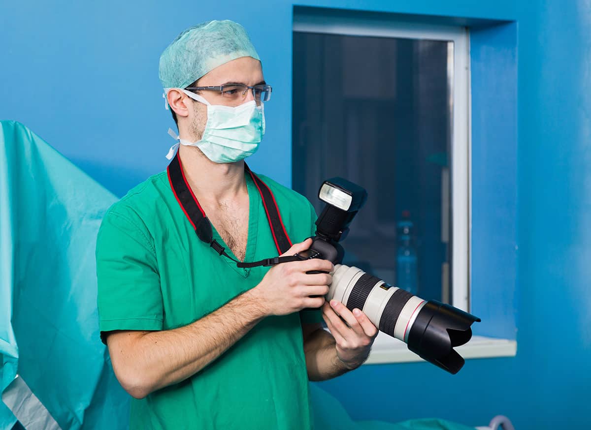
Biomedical photographers, also known as medical photographers, take detailed pictures or videos of surgical procedures, hospital patients or autopsies for medical and scientific purposes. These photographers use a variety of camera and printing techniques that allow them to create images of objects that aren’t normally visible to the human eye.
Where can a biomedical photographer work? Biomedical photographers can work in the following areas:
- Hospitals
- Pharmacy companies
- Medical offices
- Ophthalmology
- Imaging
- Health organizations
- Research facilities
- Colleges
- Medical imaging firms
Biomedical photography plays a huge role in supporting doctors and healthcare professionals. It helps them make a diagnosis and treat patients. It can also serve as instructional aids for teaching. There is a high need for biomedical photography, and because of that high need, photographers have to go through training or have training in medicine-related fields.
Table of Contents
Use of Biomedical Photography
There are three main uses of biomedical photography:
- Clinical
- Academic
- Research
Clinical
Medical photography is primarily used to record detailed and accurate images that assist doctors in diagnosing and monitoring patient’s conditions and progress once treatment starts. The photographs are an important part of the patient’s clinical notes.
Having images as documentation can help with legal aspects as well. This is to avoid allegations about improper diagnosis and inadequate surgery. Accuracy of size, position, brightness, and contrast are vital to make a conclusion while comparing preoperative and postoperative photos of results.
Academic
There is great value in using medical photographs of real patients in order to train medical students. Standard practice is to obtain permission from the patient in order to use their images for teaching purposes.
The logical justifications in the use of patient images and/or videos are:
- There is a clear educational benefit from the images in order for students to get a visual of a condition being treated and how the condition looks after treatment. It shows them what to look out for.
- Getting this type of training is beneficial to society because it allows improvement of medical care by helping to train others on disease awareness.
- Viewing images can help students test out their knowledge of what to look for in certain conditions. It gives them a chance to recognize the condition and view what it looks like when treated.
Research
Publications, thesis, and dissertations all require photographs or video clips to provide evidence that their research is of value. Being able to take different enhanced photographs of specimens such as bacteria, allows students to see more clearly and be able to conduct better research.
Conflict of Commercial Purposes
The non-medical use of images for commercial purposes can be viewed as an issue morally, ethically, and procedurally. As some people think that there is an educational value of images to society, some people argue that photographs should be of professional models. But this conflict is against showing students true results of surgery.
Here are some ethical arguments against the use of recorded images of patients for commercial purposes:
- Some patients are unable to consent prior to recording due to medical condition or emotional distress.
- Some patients feel coerced into signing consent.
- The presence of a photographer may make the patient feel embarrassed.
Fields That Rely Heavily on Biomedical Photography
Biomedical photography is used a lot in the healthcare field. These include:
- Dermatology
- Ophthalmology
- Surgery/surgical
- Oncology
- Forensics are the primary users
Dermatology
Dermatology is the branch of medicine that specializes in treating conditions of the skin, hair, and nails.
So when a patient goes to see a dermatologist for something like acne, hair loss, or psoriasis, the dermatologist will take a photo to document the initial presentation of the disease. They will then document any progress made during the treatment followed by another photo at the end of the treatment to document the before and after to their patients as well as a document that shows off the effectiveness of treatment.
An archive of documentation of a doctor’s work could be used in demonstrating treatments to show other patients or be used as marketing and promotional material.
Ophthalmology
Ophthalmology is the branch of medicine that specializes in treating conditions of the eye. Biomedical photography plays a big role in ophthalmology by using highly specialized equipment to document parts of the eye like the retina, cornea, and iris.
Special equipment is used for different parts of the eye. Something called a fundus camera can document images through the retina and cornea and let us view diseases in the eye that we normally wouldn’t be able to see.
Another type of photography that is used with the fundus camera is called Fluorescein Angiography, and that is when a fluorescent dye is injected into the bloodstream. The fluorescent dye shows up in photos of the retina and helps to differentiate retinal diseases.
A microscope and illumination device are used to photograph the cornea. These photos provide high magnification to view disorders that are impossible to see with the naked eye.
Surgery/Surgical Oncology
Surgery is the treatment of disorders or diseases by incision, while surgical oncology is the use of surgery that removes cancer. The photography can help document the growth of tumors and infection sites. It is used to communicate surgical infections, new treatment of diseases, and any toxicities of treatment.
Photography is used pre and post-operation for plastic surgery or reconstructive surgery to show a before and after of any changes made. It is used to document wound and scar healing after an incision. Plastic surgeons also use the photographs, with patient permission, to showcase their surgical abilities.
For patients undergoing radiation treatment, photography has been incorporated into treatment visits to document the progress of visible tumors.
Biomedical photography comes in handy when a clinician is not present in a patient’s appointment and is brought in to make an assessment for treatment.
Forensics
Forensics provides insight into the cause of death and clues that help law enforcement solve crimes. Photographs before and after an autopsy become evidence that supports an investigation.
Depending on the complexity of the case and department standards, photographers may need to capture as much as 200 photos. The photos are identified by using case numbers and are critical to an investigations’ integrity.
Becoming a photographer for forensics requires AT LEAST an Associates degree and one year of photography experience.
Becoming a Biomedical Photographer
Biomedical photographers need to be able to communicate visually and have advanced knowledge of photography equipment like lighting, lenses, tripods, and photo printing. These photographers need to be able to handle different environments and go through training.
Gain Experience
Before entering the biomedical photography field, it’s beneficial to become experienced in general photography. It is important to practice with a professional camera taking photos at different angles and working with different lighting situations as well as practice editing photos on computer software.
There are courses that are available that cover topics such as digital photography, computer software, lighting, camera effects, photo printing, and visual communication. These courses may also demonstrate uses of photography equipment like tripods, artificial lighting equipment, and lenses.
Photographers are required to have formal training in biology or medicine, along with formal training in photography. A training in anatomy and physiology can also be very useful.
Earn a Degree in Biomedical Photography
Scientific and medical photographers usually need to earn a Bachelor’s degree or an Associate’s degree in photography or medical illustrations in order to gain employment. Degree programs in biomedical photography train students to take high-resolution photographs of human or animal anatomy and physiology, mainly during a medical procedure.
These programs teach students how to translate basic knowledge of anatomy, physiology, and biological processes into clear, compelling visual images without bias.
Get Trained on Specialized Equipment
Taking photographs of anatomy often require specialized equipment. Photographers must know how to set up in operating rooms, research laboratories, and medical examiner facilities so they need to be aware of how to set everything up and where equipment should be placed.
It is important photographers know their equipment and are aware of their surroundings so that they don’t get in the way of the performing doctor and practitioners.
These photographers need to be trained in using multiple camera adjustments in order to create high-speed, microscopic images and/or time-lapse images.
Learn About Safety and Security Guidelines
Photographers who work with living patients or forensic evidence must follow certain rules and guidelines. They must have knowledge of confidentiality laws that protect patients’ rights and identity. Some safety guidelines require photography equipment to be sterilized so that it doesn’t introduce contaminants into a sterile hospital and medical environments.
Photographers need to know how to take photographs without interfering with the duties of the medical staff. For example, it could be life-threatening if a photographer accidentally bumped into a surgeon with their equipment while they were performing surgery.
Choose a Biomedical Specialty
Some programs offer a concentration in these fields that provide a more understanding of what kind of images that need to be captured. There are different types of specialties that a biomedical photographer can choose; here are some examples:
- Forensic photographers would work on crime scenes, in morgues, and photograph autopsies.
- Plastic surgery photographers will take a photo of the patient before their surgery to compare it to a photo taken after the surgery and a photo at the end of the recovery. They may sometimes take a photo during the procedure to show the patient what the doctor worked on.
- Scientific researching photographers will use special equipment that enhances a photograph to make what is being viewed visible to the naked eye. This would be a picture of blood cells, bacteria, and other microscopic mechanisms.
- Human studies photographers will take photos of the anatomy and physiology of patients to be examined and studied.
Photographers are not required to choose a field of specialty, but it is beneficial as it can increase job opportunities.
Medical Photography Equipment
It is important to have the proper photography equipment in order to capture what is needed. That equipment is:
- Digital SLR Camera
- Range of standard and macro lenses
- Lighting equipment
- Reflectors
- Tripod
Depending on the hospital needs, more specialized equipment is needed like:
- Large-format camera
- Endoscopic camera
- Ophthalmic cameras
- Scanners
- Photomicroscopes
Required Skills
Several skills are required to be a biomedical photographer. Here are a few:
- Possesses good knowledge of basic photography skills, digital photography, processing slides, and special films. Should be able to use various photographic techniques as different skills may be needed in different settings of the hospital.
- A reasonable knowledge of medical terminology and the manifestations of disease and hospital protocol.
- An interest in medicine and science.
- Strong computer skills for storage, indexing, retrieval, and distribution of images.
- Database management skills as a large inventory of images would have to be maintained, archived, and linked to patient data.
- Specialized photography skills like infrared imaging, fluorescent imaging, endoscopy, and photomicrography.
- Knowledge of projectors and screens, as well as sound systems as some hospitals, require the medical photographer to manage audiovisual presentations.
- Knowledge of medical ethics and laws of confidentiality because the photograph has to deal with highly classified information.
Soft Skills
A soft skill is a skill that isn’t required but is a good skill to have in order to interact effectively with other people.
Sensitivity
A medical photographer should be sensitive to the patient’s conditions and should be able to respect the dignity of the patient being photographed. They need to be able to make the patient feel comfortable during the session. The photographer shouldn’t say anything to the patient that may make them feel insecure and uncomfortable about their condition.
Strong Stomach
The term “strong stomach” means to be able to see or do unpleasant things without feeling sick or squeamish. Medical photographers will work in operation rooms, trauma wards, and post-mortem rooms, so they have to be prepared to see some traumatic scenes.
Medical Terms
It is good to understand basic anatomy and physiology terminology along with medical terms to understand a doctor’s language in order to be able to converse with them and capture what they are looking for.
Ideal Practice of a Biomedical Photographer
Most patients are willing to consent to let their pictures to be taken and used in presentations or for educational purposes. But some patients may not be willing to allow their photos to be used for public viewing in magazines or posters at the clinic’s office.
Archives on computers should be maintained under two broad categories:
- One where consent to the public display of pictures has been obtained
- and another where the patients have not consented.
The ideal practice of a photographer would be:
- Getting informed consent for the use of images and purpose must always be obtained from the patient or guardian.
- Specific and informed consent for photography should always be granted before taking photographs.
- Consent may be withdrawn at any time.
- Photography by a doctor or trained medical photographer in a suitable environment is best.
- During the photography session, the rights and dignity of the patient should always be respected.
- Images must be stored in a safe environment.
- Recognizable tattoos or birthmarks should be avoided, and eyes should only be included when absolutely necessary.
In rare cases, a patient’s consent may not be obtained in situations like filming trauma resuscitations for improving education. A patient’s confidentiality must still be respected, and images should be limited to medical record documentation. Once patient is stabilized, consent for images must be obtained, but if the patient refused consent, images must be deleted.
How to Photograph a Patient
Photographers typically will work in a hospital or inside a clinic with a slew of different professionals like doctors, nurses, care specialists, and researchers. Sometimes photographs can be delicate because of the illnesses or injuries, but there are still protocols that need to be followed.
Patients should be free of jewelry and makeup so that there aren’t any distractions in the photographs. Long hair should be pulled back, so it’s out of the face. The pose should be straightforward with feet hip-width apart.
A patient’s pose should be simple because biomedical photography seeks to show pathology, and any distractions can show an inaccurate representation.
History of Biomedical Photography
The first application of photography to medicine appeared in 1840 when Alfred Donné photographed sections of teeth, bones, and red blood cells using an instrument called microscope daguerreotype.
In 1983, the first medical photography book called “La Photographie Medicale” was published in France. Once there was improvement in photographic technique, photographs began to replace illustrations in medical books.
Soon doctors began to use photographs to document clinical cases to aid diagnosis.
Now today, hospitals hire a medical photographer whose purpose is solely to photograph a patient’s medical conditions and archive those photos into a searchable database.
Conclusion
Biomedical photography is a unique form of photography because it isn’t used as an artistic form. This doesn’t take away from its importance, however. This specialty revolves more around having an interest in a very technical form of photography that is used for the medical profession.

